
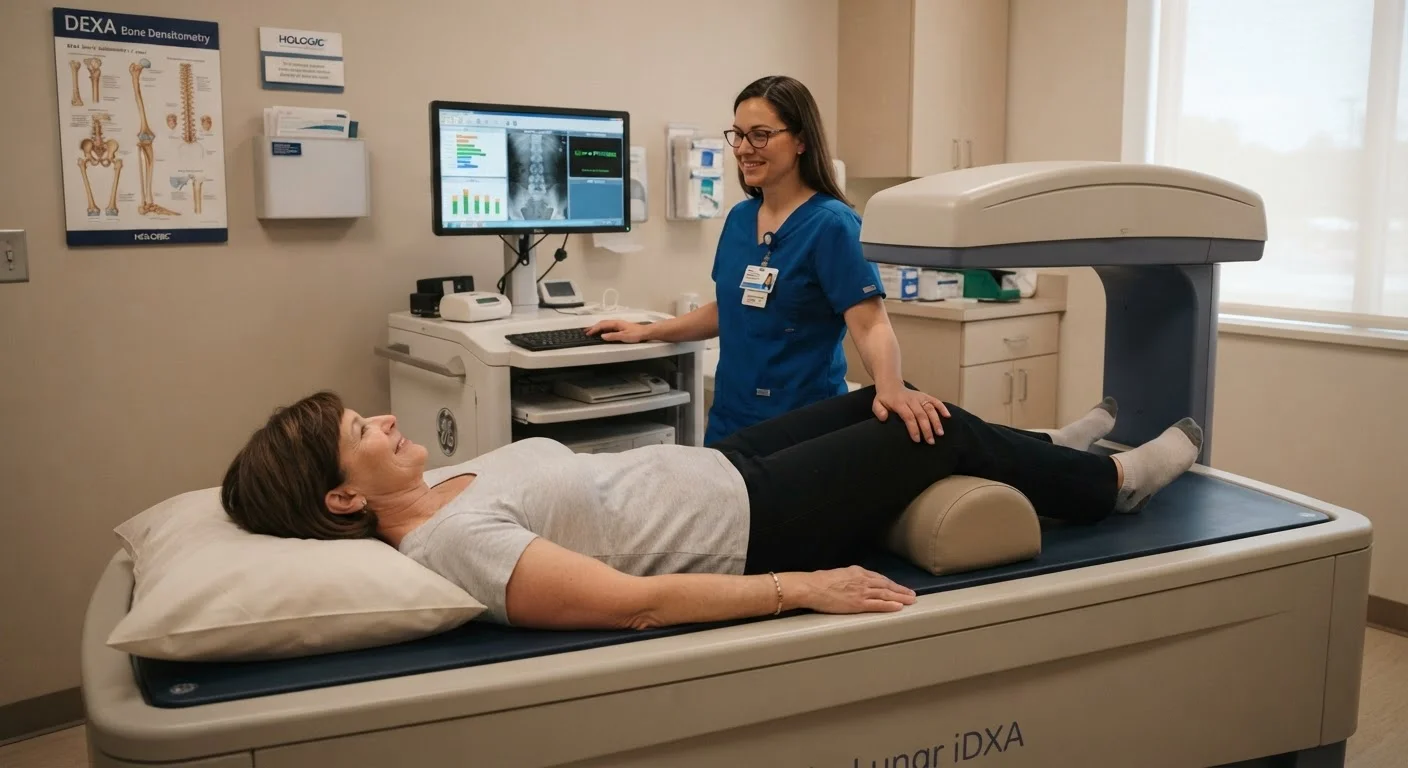
What a DEXA Scan Actually Measures
When a doctor orders a DEXA scan, they are asking one focused question: how much mineral is packed into your bones? Bone mineral density (BMD) reflects the concentration of calcium and other minerals within bone tissue. The denser the bone, the stronger and more resistant to fracture it tends to be.
The scan focuses most often on two sites: the lumbar spine (lower back) and the hip — specifically the femoral neck, where fractures in older women are most consequential. Sometimes the forearm is also measured. These sites are chosen because they represent areas most vulnerable to osteoporosis-related fractures.
The result is expressed as a number in grams per square centimeter, but what most people see reported are two derived scores: the T-score and the Z-score.
DEXA Is Different From Other Bone Tests
A standard X-ray can detect bone loss, but only after roughly 30% of density has already been lost — making it a poor early-detection tool. Blood and urine tests for bone turnover markers exist but measure the rate of bone breakdown rather than current density. DEXA is specifically designed to quantify bone mineral content with precision, which is why it remains the clinical standard for diagnosis and monitoring.
Understanding Your T-Score and Z-Score
The T-score compares your bone density to the average peak bone mass of a healthy 30-year-old adult of the same sex. This is the number most relevant to fracture risk and diagnosis:
- -1.0 and above: Normal bone density
- Between -1.0 and -2.5: Osteopenia (lower than average, but not yet osteoporosis)
- -2.5 or below: Osteoporosis
The Z-score compares your density to others of your same age, sex, and body size. A Z-score below -2.0 suggests your bone loss may be greater than expected for your age group, which can prompt your doctor to investigate secondary causes such as medication side effects or other medical conditions.
To understand how these diagnoses differ in scope and treatment implications, see our article on osteoporosis vs. osteopenia.
1 in 2
Women over 50 affected by osteoporosis-related fracture
According to the National Osteoporosis Foundation, approximately one in two women over age 50 will experience a fracture related to low bone density in their lifetime.
~30 min
Typical duration of a full DEXA scan
A complete DEXA scan of the spine and hip typically takes between 10 and 30 minutes, with no recovery time required afterward.
65+
Recommended screening age for average-risk women
The U.S. Preventive Services Task Force recommends bone density screening for all women aged 65 and older, regardless of symptoms.
Who Should Get Screened, and When
The U.S. Preventive Services Task Force recommends bone density screening for all women aged 65 and older. Younger postmenopausal women may be referred earlier if they carry specific risk factors, including:
- Low body weight or a small frame
- Early or surgical menopause
- Long-term use of corticosteroids or certain other medications
- A personal or family history of fractures
- Smoking or heavy alcohol use
If any of these apply to you, it is worth raising the topic at your next appointment. Screening is a proactive step — not a response to symptoms, since bone loss typically occurs without pain or visible signs until a fracture happens.
Prepare for Your Appointment
On the day of your DEXA scan, avoid taking calcium supplements for at least 24 hours beforehand, as they can interfere with the reading. Wear comfortable clothing without metal zippers or clasps, and let the technologist know if you have had any recent contrast imaging studies or bone scans. No fasting or special preparation is otherwise required.
How Results Fit Into Your Broader Health Plan
A DEXA result is a data point, not a verdict. Your provider will interpret it alongside your age, fracture history, medications, and other health factors. Tools like the FRAX calculator — developed by the World Health Organization — combine BMD with clinical risk factors to estimate your 10-year probability of a major fracture, giving a fuller picture than the T-score alone.
If your scan reveals low density, there is meaningful action you can take. Nutrition plays a direct role: calcium and vitamin D support bone mineral maintenance, and a well-rounded diet matters beyond just those two nutrients. You can explore practical dietary approaches in our guide on eating for bone health.
Physical activity also plays a measurable role. Not all exercise builds bone equally — our article on weight-bearing exercise and bone strength explains which types of movement are most effective.
Medical treatments for low bone density also exist and may be appropriate for some women; your doctor is the right person to discuss whether those options suit your situation.
This article is for general informational purposes only and is not a substitute for professional medical advice, diagnosis, or treatment. Always consult a qualified healthcare provider with questions about your bone health or any medical condition.